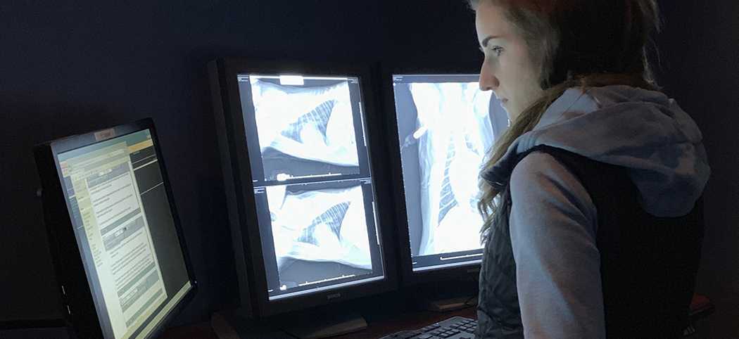
OVC Graduate Student Prototypes Equine 3D Teaching Model
November 08, 2021
Graduate students at the University of Guelph’s Ontario Veterinary College (OVC) are exploring, studying and advancing knowledge in One Health, veterinary and human health-related sciences.
Across OVC’s four academic departments, these students are exploring translational and comparative research in fields including cancer, cardiovascular disease and neuroscience, infectious diseases and immunology, epidemiology and public health, animal health management and welfare, antimicrobial resistance and stewardship, stem cell biology and regenerative medicine, and reproductive biology.
We’re highlighting graduate student work at OVC. Read on to learn more.

Alexandra Beaulieu, DVSc Student, Department of Clinical Studies
How did you find out about Clinical Studies graduate programs, and what made you want to complete your DVSc at the OVC?
I was introduced to the Clinical Studies graduate programs and Doctor of Veterinary Science (DVSc) degree in 2016, when I initially applied to complete my residency in Diagnostic Imaging at the Ontario Veterinary College (OVC). I then met my mentor Dr. Alex zur Linden, Associate Professor in Diagnostic Imaging from OVC’s Department of Clinical Studies. His passion for research and teaching combined with the innovative nature of the project captured my interest.
What is the main focus of your research?
The focus of my research was to implement simulation-based medical education in veterinary medicine to teach ultrasound-guided injections of the equine cervical articular process joints. Articular process joints are paired joints uniting adjacent vertebrae in the cervical, thoracic and lumbar spine.
The first objective of the project was to compare the visual (qualitative) ultrasonographic characteristics of 3D printed models of an equine cervical articular process joint to that of a dissected equine cervical spine.
The second objective was to develop, build and validate a 3D printed model of an equine cervical articular process joint embedded in ballistic gelatin (a dense gelatin which simulates the soft tissues surrounding the bones) to teach ultrasound-guided intra-articular injections, injections delivered to treat conditions such as osteoarthritis.
What is an anatomical 3D printed equine joint model, and why is it useful for training of ultrasound-guided injections?
In brief, 3D printing is a prototyping process where a real object is created from a 3D design. For my research project, we replicated the cervical articular process joint of a horse by using computed tomographic (CT) images of an equine neck. With the help of John Phillips from the Interdisciplinary Design Laboratory at the University of Guelph College of Arts, we transformed 2D CT scan images into 3D models. Following 3D printing, the models were assembled and embedded using ballistic gelatin.
Simulation-based medical education using 3D printed models for teaching purposes is an ethical and effective approach in healthcare education that helps to develop cognitive and technical skills and allows students to practice precise needle placement required in ultrasound-guided injections.
Why is this research important to you? Why are you passionate about this field of study?
It is remarkable to be able to create a teaching tool that simultaneously provides an environment that closely approximates the characteristics of a clinical situation, mimics the visuospatial and real-time aspects of a procedure, provides realistic tactile feedback, and allows an objective assessment of the performance under study.
Why is there a need for this research?
While the efficacy of intra-articular injections of glucocorticoids to treat osteoarthritis of the equine cervical articular process joints is well recognized, this invasive procedure is not without risks. It is therefore essential that equine veterinarians receive training to practice and refine this technique to minimize risks for complications during the procedure.
Considering the proven benefits of simulation-based medical education, the development and validation of an equine cervical articular process joint model to teach ultrasound-guided injections and treat horses suffering from cervical osteoarthritis was lacking in the current literature.
In plain language, how would you describe the benefits of your research?
We hope this new simulation-based training model will provide fourth-year veterinary students, equine practitioners, and veterinary specialists the opportunity to develop and improve their skills and confidence to perform cervical ultrasound-guided articular process joint injections while minimizing procedural complications and ensuring quality care for patients.
Who is your advisor(s) and who else is affiliated with your research?
My primary advisor is Dr Alex zur Linden, OVC Department of Clinical Studies, and my advisory committee includes Drs. Stephanie Nykamp, OVC Associate Dean, Clinical Program, John Phillips, Interdisciplinary Design Laboratory, College of Arts, Luis Arroyo and Judith Koenig, OVC Department of Clinical Studies
Who are your current funders for this research?
My research project was funded by grants from Equine Guelph, (EG201704), and SONAMI.
Can you provide any links to published journals for these projects? Is there any research in progress that is expected to be released soon?
Merchan A, Beaulieu A, Côté N, zur Linden A. Use of computed tomography angiography for the evaluation of a hemangioma in a Standardbred horse. Equine Vet Educ.
-
Accepted, to be included in a volume.
Lee GKC, Bowal J, Beaulieu A, Iverson M, Jensen M, Mutsaers T, Foster RA, Wood RD. Alveolar rhabdomyosarcoma affecting the head of a young dog. J Am Vet Med Assoc.
-
Accepted, to be included in a volume.
Beaulieu A, Nykamp S, Phillips J, Arroyo LG, Koenig J, zur Linden A. Development and validation of a three-dimensional printed training model to teach ultrasound-guided injections of the cervical articular process joints in horses. J Vet Med Educ.
-
https://pubmed.ncbi.nlm.nih.gov/34115577/
Beaulieu A, Stachewski Zakia L, Ludwig L, Kenney D, Rätsep E, Nykamp S. Oesophageal muscular hypertrophy and pulsion diverticulum in a Friesian horse. Vet Rec Case Rep. 2020;8(3):1-4.
Beaulieu A, zur Linden A, Phillips J, Arroyo LG, Koenig J, Monteith G. Various 3D printed materials mimic bone ultrasonographically: 3D printed models of the equine cervical articular process joints as a simulator for ultrasound guided intra-articular injections. PLoS One. 2019;14(8):e0220332.
Slater RT, Beaulieu A, Schachtel J, Wilkinson TE. What Is Your Diagnosis? Perforating esophageal foreign body in a dog. J Am Vet Med Assoc. 2019;255(4):423-5.
-
https://avmajournals.avma.org/doi/abs/10.2460/javma.255.4.423?journalCode=javma
.png)